Download Online From Protein Structure
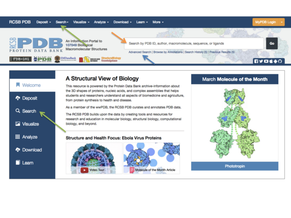
As a member of the wwpdb the rcsb pdb curates and annotates pdb data according to agreed upon standards.

Download online from protein structure. A protein accession number eg. The pdb protein data bank is the largest protein structure resource available online. Polypeptide sequences can be obtained from nucleic acid sequences. This list of protein structure prediction software summarizes commonly used software tools in protein structure prediction including homology modeling protein threading ab initio methods secondary structure prediction and transmembrane helix and signal peptide prediction.
Sites are offered for calculating and displaying the 3 d structure of oligosaccharides and proteins. O all functional proteins will have up to 3 tertiary level of structures. This is done in an elegant fashion by forming secondary structure elements the two most common secondary structure elements are alpha helices and beta sheets formed by repeating amino acids with the same fps angles. Users can perform simple and advanced searches based on annotations relating to sequence.
The pdb archive contains information about experimentally determined structures of proteins nucleic acids and complex assemblies. The rcsb pdb also provides a variety of tools and resources. Protein mixtures can be fractionated by chromatography. 24 primary structure of proteins the amino acid sequence or primary structure of a purified protein can be determined.
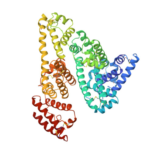
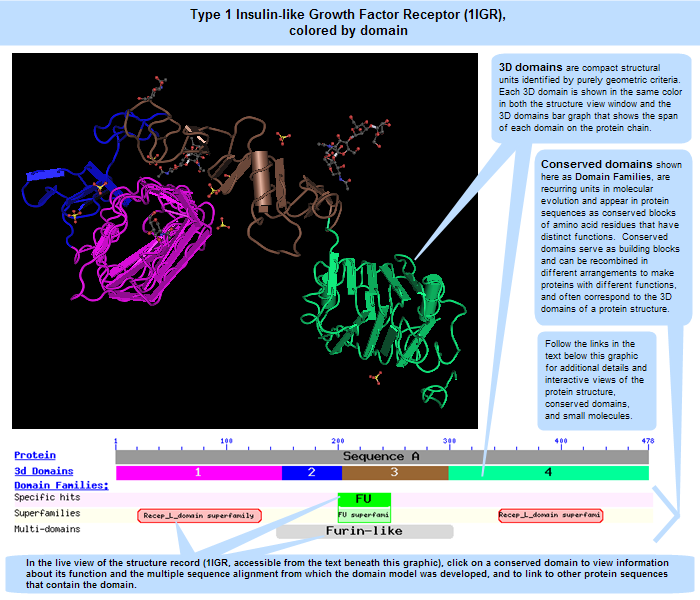
Secondary structure the primary sequence or main chain of the protein must organize itself to form a compact structure. Use the finding a structural template guide to find the most appropriate pdb. The pdb archive contains information about experimentally determined structures of proteins nucleic acids and complex assemblies. The 3d view of the structure you have uploaded will now be displayed.
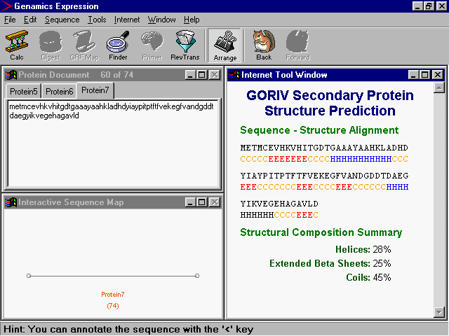
In order to load the pdb type the below command from biopdb import protein structure file formats. Secondary structure refers to the coiling or folding of a polypeptide chain that gives the protein its 3 d shapethere are two types of secondary structures observed in proteins. With the two protein analysis sites the query protein is compared with existing protein structures as revealed through homology analysis. Select the file and click load.
As a member of the wwpdb the rcsb pdb curates and annotates pdb data according to agreed upon standards. One type is the alpha a helix structurethis structure resembles a coiled spring and is secured by hydrogen bonding in the polypeptide chain. Try out the new interactive 3d structure viewer icn3d. It hosts a lot of distinct protein structures including protein protein protein dna protein rna complexes.
The rcsb pdb also provides a variety of tools and resources. O some proteins will have all the 4 levels of structures up to quaternary structure. Proteins and other charged biological polymers migrate in an electric field. O primary structure of a protein gives the details of the amino acid sequence of a.

